cardiac Biopsy
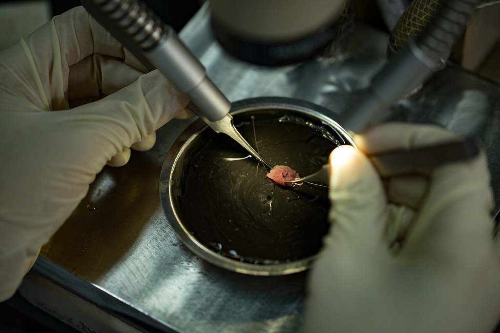
Overview: In a myocardial biopsy, doctors remove a small piece of heart tissue and examine it. It is usually done after transplants to see how your body is reacting to your new heart.
A myocardial biopsy is a procedure in which a small piece of the heart muscle is removed for laboratory examination. Biopsies are routinely performed after heart transplants to watch for signs of rejection. They are also done if you have signs of myocarditis, cardiac amyloidosis, or certain cardiomyopathies other than HCM. Most HCM patients never have a biopsy.
The doctor will give you instructions about how to prepare for the biopsy. Usually these include not eating or drinking anything for 6-8 hours before the test. Before the procedure, you will be given a sedative to relax but you will remain awake. You will lie down flat on a table during the exam and will be able to follow instructions during it. Your skin will be scrubbed with a local anesthetic and a surgical cut will be made into the area where the catheter will be inserted (arm, neck, or groin). The catheter is then inserted into a vein or artery, depending on whether tissue will be collected from the right or left side of your heart. Once the catheter is guided through the vessel using x-ray imaging and is in position, a device will remove a small piece of tissue.
The risks of biopsies include blood clots, bleeding from the puncture site, arrhythmias, infection, injury to a vein or artery, pneumothorax, and very rarely ruptured heart.
Your doctor will discuss the results with you. Sometimes abnormal tissue is in the heart but is missed by this procedure.
MedlinePlus. (2020, October 8). Myocardial biopsy. Myocardial biopsy: MedlinePlus Medical Encyclopedia. Retrieved October 17, 2020, from https://medlineplus.gov/ency/article/003873.htm
HCMA 6/2021


